

Corrective facial procedures increasingly rely on biomedical engineering to improve anatomical accuracy, functional preservation, and long term outcome stability. Modern corrective interventions often involve congenital abnormalities, traumatic injuries, structural airway issues, postoperative revisions, and asymmetry management. These cases require coordinated planning between reconstructive surgery, imaging science, biomaterials engineering, biomechanics, and regenerative medicine. The integration of engineering systems into facial surgery has shifted many procedures from estimation based approaches toward protocol driven planning supported by measurable anatomical data.
Advanced Imaging Systems in Corrective Facial Assessment
High resolution imaging remains one of the most important biomedical contributions to corrective facial work. Traditional examination methods provide surface level observations, while imaging technologies generate detailed internal maps of bone, cartilage, soft tissue layers, and airway structures.
Common diagnostic systems include:
- Computed tomography for skeletal evaluation
- Magnetic resonance imaging for soft tissue analysis
- Three dimensional facial scanning for symmetry assessment
- Digital surface mapping for contour analysis
- Computer assisted reconstruction software for surgical simulation
These technologies support preoperative planning by identifying anatomical variations before intervention begins. Cause and effect relationships become easier to predict because surgeons can evaluate how structural adjustments may influence neighbouring tissues.
For example, alteration of nasal support mechanisms may affect airflow resistance, soft tissue projection, and facial balance. Similarly, midface reconstruction may influence orbital positioning and cheek contour distribution.
Three Dimensional Modelling and Symmetry Optimization
Biomedical engineering uses digital modelling systems to convert imaging information into measurable anatomical representations. Three dimensional analysis improves precision by allowing comparison between existing structures and projected corrections.
Parameters commonly evaluated include:
- Nasal projection angles
- Septal alignment
- Midfacial width ratios
- Soft tissue thickness
- Orbital relationships
- Jaw positioning
- Airway dimensions
This process reduces variability during surgical planning and supports objective decision making.
Practical application becomes particularly important in post traumatic reconstruction. Patients with previous fractures may demonstrate irregular healing patterns that affect symmetry and function. Digital modelling assists in identifying displacement zones and estimating corrective requirements before surgery.
Biomechanics and Functional Preservation in Nasal Revision Cases
Revision procedures represent a clinically demanding category because tissue architecture may already be altered by previous intervention, scar formation, cartilage depletion, or structural instability. Biomedical engineering contributes through biomechanical analysis that evaluates tissue behaviour under functional loads.
Areas commonly assessed include:
- Airflow dynamics
- Cartilage stability
- Pressure distribution
- Skin elasticity response
- Structural load transfer
- Internal valve integrity
Clinical planning frequently involves combining imaging findings with biomechanical models to reduce postoperative complications.
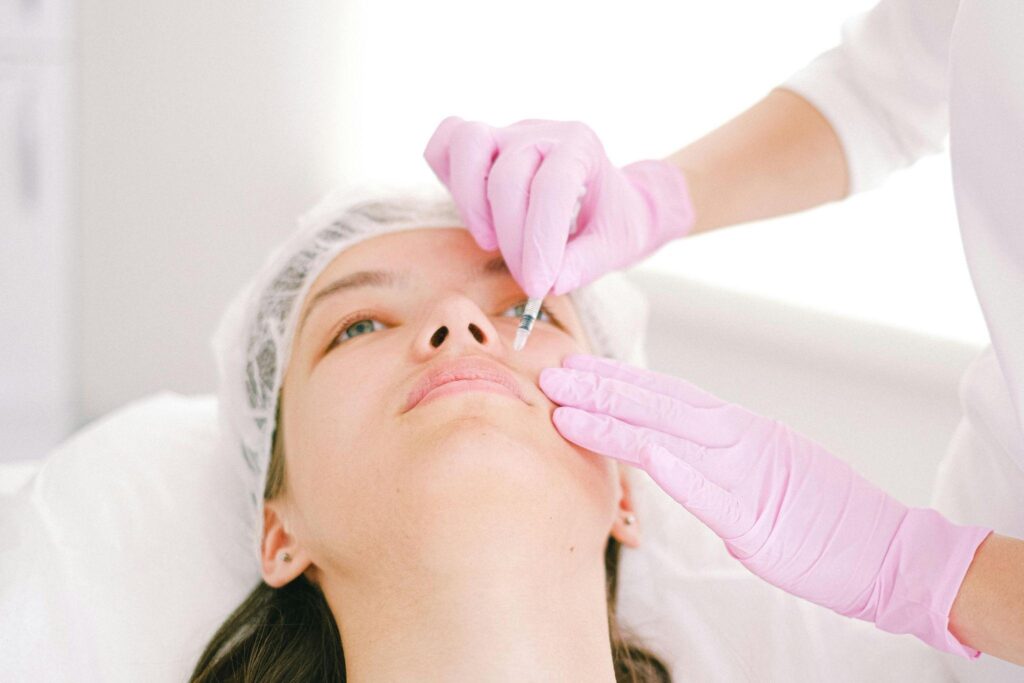
A practical example can be observed in patients undergoing secondary nasal correction after previous cosmetic or functional procedures. Cases requiring cartilage support restoration often benefit from detailed anatomical evaluation similar to approaches used in revision rhinoplasty procedures performed in Glendale for complex structural correction, where preservation of breathing function remains equally important as aesthetic refinement.
This type of protocol driven assessment helps optimize outcomes while reducing risks associated with collapse, asymmetry, or airway compromise.
Tissue Engineering and Regenerative Support Systems
Regenerative medicine has expanded corrective possibilities by introducing biologically active materials intended to support tissue repair and reconstruction.
Biomedical applications currently include:
- Cellular scaffolds
- Biologic matrices
- Engineered cartilage frameworks
- Growth factor delivery systems
- Tissue regeneration platforms
Traditional reconstruction often depends on graft harvesting from existing tissue sources. Tissue engineering attempts to improve repair environments by promoting regeneration and improving integration.
Clinical relevance may include:
Cartilage Reconstruction
Patients presenting with traumatic loss, congenital deficiency, or previous surgical reduction may require structural reinforcement. Engineered materials provide additional options for support restoration.
Soft Tissue Defect Management
Volume deficiencies resulting from injury or postoperative changes can benefit from regenerative approaches aimed at improving tissue quality.
Scar Control and Healing Optimization
Biologic scaffolds may contribute to organized healing patterns by supporting tissue stability during recovery.
These interventions remain under continuous study, but current evidence indicates increasing value in reconstructive planning.
Computer Assisted Navigation and Procedural Accuracy
Facial surgery involves narrow anatomical margins where millimetre scale deviations may influence outcome quality. Computer assisted navigation systems improve procedural control by matching surgical activity with digital imaging references.
Functions supported by navigation technology include:
- Identification of anatomical landmarks
- Alignment verification
- Implant positioning control
- Intraoperative orientation
- Risk reduction near critical structures
Applications frequently extend to:
- Midface reconstruction
- Orbital repair
- Craniofacial correction
- Secondary revision procedures
- Complex asymmetry management
Cause and effect relationships become particularly relevant because improved alignment accuracy may reduce revision rates and improve structural stability.
Artificial Intelligence and Predictive Outcome Analysis
Artificial intelligence supports biomedical engineering by analysing large datasets related to anatomy, recovery patterns, and procedural outcomes.
Potential clinical functions include:
- Recovery prediction models
- Symmetry analysis
- Tissue response monitoring
- Risk stratification systems
- Surgical simulation support
AI systems remain supportive rather than independent decision makers. Their primary value lies in organizing complex information and assisting clinicians during planning stages.
Outcome optimization depends on integrating these systems with clinical judgement, imaging interpretation, and reconstructive protocols.
Conclusion
Biomedical engineering has become a central component in corrective facial procedures through its influence on imaging, modelling, biomechanics, regenerative medicine, navigation systems, and predictive technologies. The transition from observational planning toward evidence supported workflows has improved anatomical precision, functional preservation, and risk management.
Cross disciplinary integration between engineering and surgery continues to strengthen outcome optimization strategies. As tissue engineering, artificial intelligence, and patient specific modelling advance further, corrective facial procedures are expected to become increasingly individualized, function focused, and supported by measurable biological data rather than visual estimation alone.
