

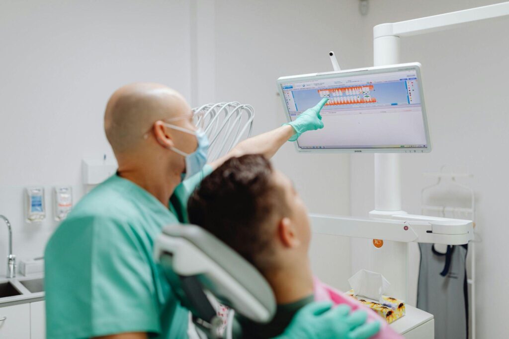
Digital smile design has shifted from a purely cosmetic workflow into a structured clinical system that combines restorative dentistry, facial biometrics, soft tissue analysis, artificial intelligence, and digital manufacturing. Current protocols emphasize treatment predictability, tissue preservation, variability reduction, and functional integration rather than isolated aesthetic modification. Modern workflows increasingly rely on facial recognition systems, intraoral scanning, occlusal mapping, and restorative simulations to improve treatment planning accuracy while reducing procedural uncertainty.
Diagnostic Mapping and Facial Recognition Integration
Advanced smile design protocols commonly begin with digital screening and facial analysis. Clinical evaluation extends beyond tooth shade and alignment and now includes facial proportions, lip support, gingival exposure, dynamic smile movement, and symmetry assessment. Biometrics and facial recognition systems help standardize measurements and reduce operator dependent variability.
Typical diagnostic parameters include:
- Smile arc position
- Midline alignment
- Gingival display at rest and during expression
- Lip mobility patterns
- Facial thirds and proportional balance
- Soft tissue support around the perioral region
- Occlusal force distribution
These measurements support restorative planning while identifying risk flags that may influence treatment sequencing. For example, excessive gingival exposure combined with inflammatory tissue changes may require periodontal stabilization before veneer placement or restorative intervention.
Soft Tissue Stability and Gingival Inflammation Control
Soft tissue health remains a major determinant of smile design outcomes. Gingival inflammation alters contour visibility, affects impression accuracy, and increases variability during digital scanning. Clinical protocols therefore prioritize tissue stabilization before aesthetic procedures.
Pre treatment screening frequently includes:
- Plaque index evaluation
- Gingival bleeding assessment
- Barrier tissue inspection
- Soft tissue thickness analysis
- Inflammation grading
- Periodontal risk documentation
Cause and effect relationships are important during planning. Inflamed gingiva may enlarge tissue dimensions and create inaccurate restorative margins. If veneer preparation occurs before inflammation resolution, final contours may become inconsistent once healing is complete.
A preventive approach often involves staged management:
- Initial periodontal evaluation
- Tissue stabilization phase
- Digital scan acquisition
- Restorative simulation
- Final preparation sequence
This sequence supports outcome optimization and reduces correction procedures.
Ultra Thin Restorations and Tissue Preservation Protocols
Modern restorative engineering favors minimally invasive approaches with emphasis on enamel preservation. Ultra thin ceramic restorations reduce structural removal while maintaining optical characteristics similar to natural enamel.
Clinical advantages include:
- Reduced preparation depth
- Improved enamel bonding surface
- Lower sensitivity risk
- Better light transmission properties
- Increased tissue preservation
In restorative planning, clinicians may compare preparation strategies according to facial proportions and tissue response. Patients with thin gingival biotypes, high smile lines, or visible cervical areas often require more conservative sequencing to avoid margin visibility and soft tissue irritation.
In some advanced cosmetic protocols, clinicians evaluating custom veneer approaches available in New York City practices may use digital smile simulations to determine whether porcelain restorations improve facial harmony without excessive enamel reduction. This step helps integrate restorative goals with biometrics and functional analysis rather than relying solely on visual preference.
Artificial Intelligence and Predictive Treatment Modeling
AI supported systems increasingly assist with diagnostic consistency and restorative design. These systems evaluate scan datasets, facial photographs, and occlusal information to improve predictive planning.
Applications include:
- Tooth proportion recommendations
- Bite stress visualization
- Margin identification support
- Functional load mapping
- Restorative longevity estimation
Practical examples are useful during treatment selection. A patient presenting with uneven wear patterns and parafunctional loading may appear suitable for aesthetic veneers initially. However, bite force mapping may reveal concentrated posterior stress requiring occlusal management before restorative placement.
This reduces fracture risk and improves long term restoration survival.
3D Manufacturing and Material Compatibility
Advanced dental engineering now integrates CAD/CAM workflows with additive manufacturing systems. Printed prototypes, mock ups, and provisional restorations support procedural verification before definitive placement.
Common materials include:
- Ceramic reinforced composites
- Hybrid resin systems
- High translucency ceramics
- Biocompatible provisional materials
Material selection depends on:
- Occlusal demand
- Tissue response
- Wear resistance needs
- Esthetic requirements
- Functional loading patterns
Material compatibility also influences post treatment adaptation. Rigid materials placed in patients with heavy functional activity may require additional monitoring to prevent stress concentration and marginal instability.
Treatment Sequencing and Recovery Monitoring
Procedure timing significantly affects smile design predictability. Simultaneous interventions may increase cumulative irritation and interfere with tissue recovery.
Recommended sequencing may involve:
Phase One: Assessment
- Facial analysis
- Digital photography
- Intraoral scanning
- Gingival evaluation
- Occlusal documentation
Phase Two: Stabilization
- Inflammation reduction
- Periodontal management
- Tissue monitoring
- Barrier recovery support
Phase Three: Restorative Planning
- Digital simulations
- Mock up approval
- Margin planning
- Material selection
Phase Four: Placement and Recovery
- Restoration delivery
- Occlusal refinement
- Tissue observation
- Functional adaptation review
Post treatment monitoring remains essential because soft tissue remodeling may continue during early recovery periods. Clinicians often document:
- Gingival contour stability
- Sensitivity response
- Margin adaptation
- Occlusal comfort
- Inflammatory changes
Early monitoring improves variability control and supports long term success.
Future Direction of Digital Smile Engineering
Digital smile design continues moving toward standardized, evidence guided workflows that combine restorative dentistry, facial biometrics, tissue physiology, and computational analysis. The objective is no longer limited to aesthetic enhancement. Modern protocols focus on reproducibility, risk reduction, tissue preservation, and functional integration.
As facial recognition systems, AI diagnostics, and restorative engineering continue to evolve, treatment planning becomes increasingly precise. Structured screening, inflammation control, conservative preparation methods, and recovery monitoring remain central components for achieving predictable smile outcomes while maintaining biological stability and long term restorative performance.
